| Dislocated total hip arthroplasty |
Left hip unipolar prosthesis (hemiarthroplasty) dislocation |
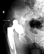
Bilateral total hip arthroplasty; right hip arthroplasty dislocation |
Metal-on-metal left hip arthroplasty with Metallosis and bony erosions |
 |
 |
 |
 |
Most hip arthroplasty dislocations are posterior. From Benjamin, 1994 |
|
Elderly woman with bilateral press fit total hip arthroplasties. There is a dislocation on the right. |
68 year-old woman with left hip metal-on-metal prosthesis. Bony erosions (arrows) are evident on the greater and lesser trochanter from probable metallosis with pseudotumor formation. |
 |
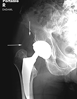
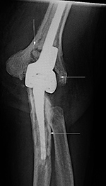
| Right hip dislocation with displaced polyethylene liner |
|
 |
 |
 |
|
| Initial AP radiograph (left) shows a right hip dislocation. Note the polyethylene liner (arrow) is still associated with dislocated femoral head. The hip was subsequently relocated. The AP radiograph (middle) show subtle eccentric positioning of the reduced femoral head within the acetabular cup. A subtle density (arrows) represents the liner which is now disassociated from the femoral head. This is somewhat more clearly shown (arrow) on the frog leg lateral view (right). |
|
|
| Stress shielding |
Stress loading |
|
 |
 |
 |
|
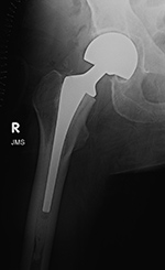
| 69 year-old woman with right femoral neck fracture treated with bipolar hip prosthesis (left image). Five months later (right image) there is reduction of femoral cortical thickness (lower two arrows) and osteopenia in the greater trochanter (top arrow) from stress shielding. |
52 year-old man with right total hip arthroplasty 7 years previously. An AP radiograph of the pelvis shows the arthroplasty with considerable thickening to lateral and medial femoral cortices from stress loading. A right iliac reconstruction plate is partially visualized. |
|
|
| Metal-on-metal right Hip arthroplasty with heterotrophic bone formation |
Right Hip Arthroplasty with heterotrophic bone formation |
Bony osteolysis |
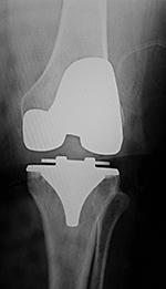
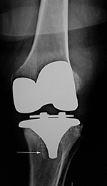
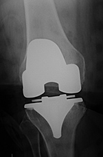
Subtle failure of total knee arthroplasty (TKA) |
 |
 |
 |
 |
| 55 year-old man with right hip metal-on-metal arthroplasty and heterotrophic bone formation above the greater trochanter. The metal-on-metal prosthesis was removed and replaced with a standard hip prosthesis due to continued hip pain and chronic fluid collection around the greater trochanter. A later scout CT image shows increased heterotrophic bone formation (arrow) near the revision prosthesis. The heterotrophic bone was later removed. |
A resurfacing hip arthroplasty is present. The polyethylene acetabular component is radiolucent. Non-infectious osteolysis (arrow) is present at the tip of the femoral stem. From Benjamin, 1994 |
A noncemented total knee arthroplasty is present with a metal backed patellar component. There is also a polyethylene locking clip which locks the tibial polyethylene into the tibial base plate. The anterior cortex of the femur is notched and eroded (top arrow). There is also subtle subsidence of the tibial component (bottom arrow). From Benjamin, 1994 |
 |
| Aseptic loosening of bilateral total knee arthroplasties |
 |
 |
 |
 |
| 65 year-old woman with bilateral cruciate-retaining total knee arthroplasties (TKAs) placed in early 2009. In November 2015 routine follow-up showed bilateral aseptic loosening in both tibial components with wide periprosthetic lucencies (arrows) and varus deformity on the left. From left to right, respectively, AP radiograph of the left knee in the postoperative period, AP radiograph of the left knee 6.5 years later, lateral radiograph of the left knee in the postoperative period, and lateral radiograph of the left knee 6.5 years later. Images courtesy Laura H Lee, MD. |
|
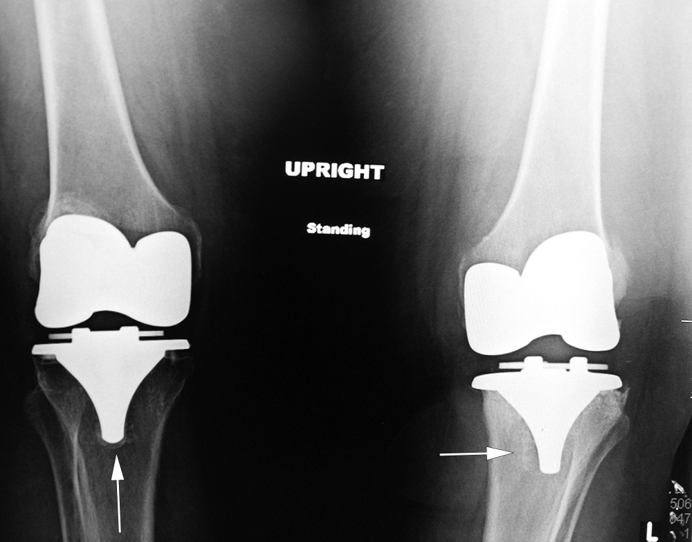
| Aseptic loosening of bilateral total knee arthroplasties continued |
 |
 |
 |
|
| 65 year-old woman with bilateral cruciate-retaining total knee arthroplasties (TKAs) placed in early 2009. In November 2015 routine follow-up showed bilateral aseptic loosening in both tibial components with wide periprosthetic lucencies (arrows) and varus deformity on the left. From left to right, respectively, AP radiograph of the right knee in the immediate postoperative period, AP radiograph of the right knee 6.5 years later, and standing frontal view of the knees 6.5 years after surgery. Images courtesy Laura H Lee, MD. |
|
| Rotating-hinge knee implant |
Rotating hinge knee implant failure |
 |
 |
 |
 |
| 69 year-old man with revision of an infected right knee prosthesis (two left panels). A rotating hinge knee implant was placed. Note the antibiotic beads about the medial aspect of the implant. The two fixation screws and wire are from prior surgery. There was implant failure with disruption of the implant six weeks later (two right panels). |
|
| Infected total elbow arthroplasty with eventual placement of a revision total elbow arthroplasty |
 |
 |
 |
 |
| 66 year old man with infected left elbow arthroplasty. The arrows point to areas of periprosthetic lucency suggesting loosening which was found to result from an infected prosthesis. The patient had the prosthesis removed and underwent multiple subsequent surgeries with placement of a revision elbow arthroplasty. |