| Secure-C Cervical Artificial Disc (Globus Medical) |
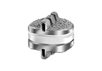
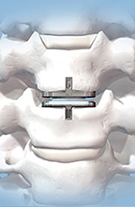
ProDisc-C Total Disc Replacement (DePuy-Synthes) |
 |
 |
 |
 |
| |
© DePuy Synthes 2016. All rights reserved. ProDisc® C is a trademark of DePuy Synthes. |
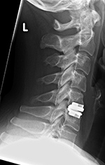
43 year-old woman with localized disk disease at C5-6 |
 |
| Mobi-C Cervical Disc (LDR Holding) |
Prestige Cervical Disc (Medtronic) |
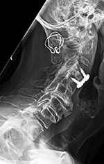
Posterior Spinal Wiring |
 |
 |
 |
 |
| |
This is a metal-on-metal artificial disk that is anchored to the vertebral bodies by anterior locking screws. Reprinted with the permission of Medtronic, Inc. © 2016 |
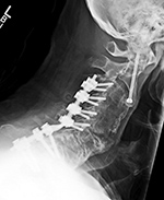
Posterior spinal wiring goes from C1 to C2. There is also solid posterior bony fusion mass from the occiput to C2 as well as an anterior cervical fusion plate and screws from C2 to C3 with interbody bony disk plug at C2-3. |
 |
| Zero-P (DePuy Synthes) low Profile anterior interbody fusion device |
Zero-P (DePuy Synthes) low Profile anterior interbody fusion device |
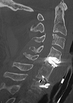
Metal-on-polyethylene disk at C4-5 and Zero-profile ACDF at C6-7. |
 |
 |
 |
 |
| © DePuy Synthes 2016. All rights reserved. ZERO-P® is a trademark of DePuy Synthes. |
ZERO-P® is a trademark of DePuy Synthes. |
61 year-old man with past trauma to the cervical spine. A sagittal CT reformatted image shows a metal-on-polyethylene disk at C4-5, bony fusion of C5-6, and a Zero-profile ACDF at C6-7. |
 |
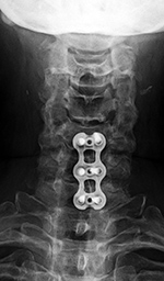
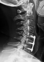
| Anterior Cervical Diskectomy and Fusion (ACDF) C5-7 |
Disk Cage |
 |
 |
 |
 |
| There are PEEK intervertebral disk cages at C5-6 and C6-7. |
These are titanium coated PEEK disk cages. |
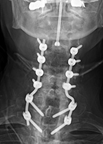
There is a hollow disk cage at C4-5 with bone chips in the cage. Posterior spinal fusion is present with lateral mass screws from C3-6 bilaterally and Pedicle screws bilaterally at T1-2 with rods running on each side from C3-T2. An anterior plate and screws is at C4-5. A laminectomy is present bilaterally through much of the cervical spine. |
 |
| Odontoid fracture Fixation |
Susceptibility artifact from cervical spine disk prosthesis |
 |
 |
 |
 |
| There is an unrelated posterior spinal fusion (PSF) at C3-T2 with lateral mass screws at C3-6 and pedicle screws at T1-2. |
|
43 year-old man with metallic cervical disk prosthesis at C5-6. Metallic susceptibility artifact completely obliterates imaging of the spine and spinal cord at this level. |
| Vertebroplasty extruded cement |
Intrathecal drug delivery catheter (arrow) |
Thoracic spinal cord neurostimulator electrodes |
Bone stimulator |
 |
 |
 |
 |
| |
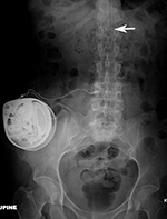
The catheter is in the lower thoracic subarachnoid space. It exits into an anterior abdominal delivery pump. From Hunter, 2004 |
|
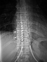
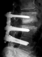
A battery pack overlies the right 12th rib. Wires are going to the bilateral bony fusion masses. There is a laminectomy from L2 to L5 with bilateral pedicle screws and a pedicle plate on the right and a connecting rod on the left. Brantigan vertebral cages are at the L5-S1 disk space. From Hunter, 2004 |
 |
| Harrington rods |
Harrington rods |
 |
 |
 |
 |
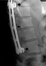
| The hooks on these rods are designed for distraction. From Hunter, 1994 |
Harrington rods are at the thorocolumbar junction stabilizing a vertebral body fracture. The hooks (thick arrows) anchor the rods in the lamina. Segmental wires around the lamina (thin arrow) supplement the fixation. From Hunter, 1994 |
Harrington rods (flanged ends) and Knodt rods (threaded rods) have hooks along the rods; designed to either distract the spine or compress it, depending on the direction in which the hooks are placed. |
 |
| Lower thoracic spine neurostimulator leads |
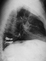
Embolization of vertebroplasty cement into the pulmonary arterial tree |
 |
 |
 |
 |
| 38 year-old woman with chronic spinal pain |
From Hunter, 2004 |
 |
| Baclofen intrathecal pump AP view |
Baclofen intrathecal pump lateral view |
Vertebroplasty at L1 and L3 (AP view) |
Vertebroplasty at L1 and L3 (lateral view) |
 |
 |
 |
 |
| The catheter (arrow) goes into the lower thoracic spinal subarachnoid space. |
The catheter (arrow) goes into the lower thoracic spinal subarachnoid space. |
There is extrusion of vertebroplasty cement into the T12-L1 disk space. |
 |
| Lumbar spine pedicle plates and screws and Brantigan disk cage (arrow) |
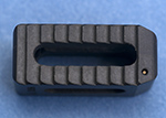
Brantigan disk cage |
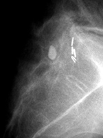
Residual Pantopaque from a distant myelogram |
 |
 |
 |
 |
| Note the associated solid posterolateral bony fusion masses |
|
From Hunter, 1994 |
|
 |
| Posterior spinal fusion (PSF) |
Lumbar spine disk spacer (artificial disk) |
 |
 |
 |
 |
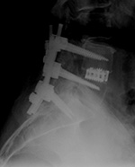
| Shown are pedicle screws and rods on each side, two crosslinks (at L4 and S1), and intervertebral disk spacers at L4-5. |
|
|
 |
| Sacral stimulator AP and lateral views |
Sacral stimulator |
Sacral stimulator |
 |
 |
 |
 |
| |
|
Hernia mesh is also visible over the lower abdomen and pelvis |
From Hunter, 2004 |
 |