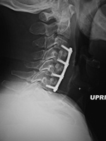
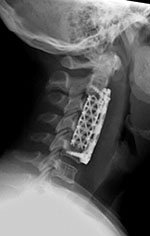
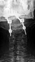
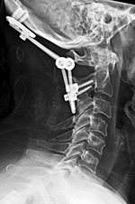
| Anterior cervical fusion plate |
Anterior Cervical Diskectomy and Fusion (ACDF) plate |
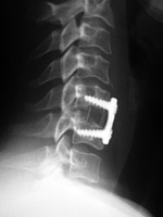
Cervical spine fusion cage and anterior cervical fixation plate |
 |
 |
 |
 |
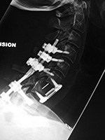
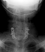
| 53 year-old man with disk herniation. Anterior cervical fusion plate spans C3 to C6. |
|
|
 |
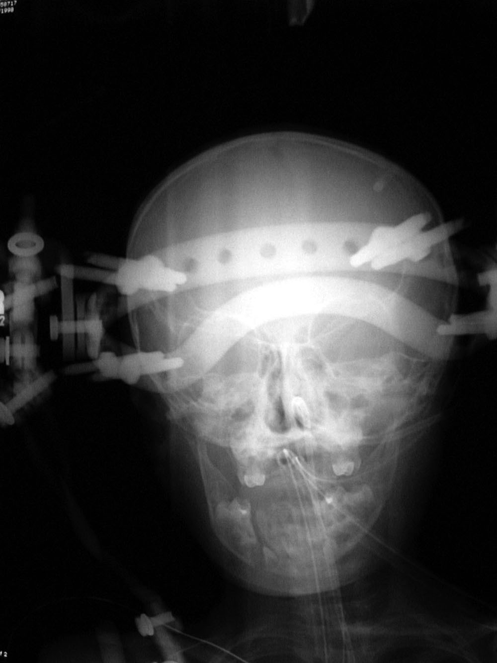
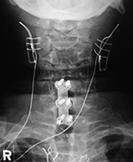
| Cranial tongs (AP view) |
Cranial tongs (lateral view) |
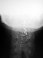
Cervical spine intervertebral disk fusion cage |
Anterior cervical disk fusion (ACDF) and PEEK cage at C5-6 |
 |
 |
 |
 |
| Child with severe intracranial and cervical spine injuries with bilateral cranial stabilization tongs, an endotracheal tube, an oroogastric tube, and a feeding tube entering via the nose.
From Hunter, 2004 |
|
|
|
| Occipital strut with posterior cervical plates |
Cervical spine anterior and posterior fusion |
Odontoid screw (nail) fracture fixation with posterior cervical fixation plates and screws |
 |
 |
 |
 |
| |
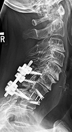
Young woman with traumatic locked facets at C6-7 and C7 body fracture. A posterior cervical fusion with lateral mass screws (cervical spine) and pedicle screws (thoracic spine) and rods extends from C4 to T2. There is an anterior cervical fusion plate and screws at C6-7 with a intervertebral disk cage at C6-7 and a crosslink at C6. |
|
|
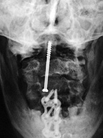
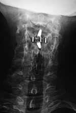
| Odontoid fixation screw AP view |
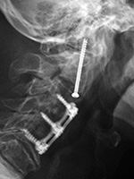
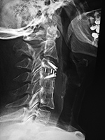
Odontoid fixation screw lateral view |
Posterior cervical spine clamp |
Odontoid fracture fixation |
 |
 |
 |
 |
| There is also an anterior cervical fusion plate and screws from C3-6. |
Patient with rheumatoid arthritis and atlantoaxial (C1-C2) subluxation and generalized cervical spine laxity |
Postoperative lateral radiograph of the cervical spine. There is fixation of an odontoid base fracture by an odontoid screw and a sublaminar wire between C1 and C2. There are also skin staples and a surgical drain in the posterior aspect of the neck |
|
| Odontoid Fracture Fixation |
 |
 |
 |
 |
| 47 year-old woman with type III dens fracture treated by occiput-C2 fusion. |
|
| Two odontoid screws |
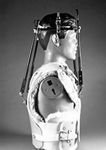
Cranial (head) tongs |
 |
 |
 |
 |
| 86 year-old woman with C1 posterior ring and type II dens fractures two months earlier treated with two odontoid (dens) screws. |
Cranial (head) tongs are used to stabilize the head and neck in a patient with a cervical spine fracture. One or more screws penetrate the outer table of the skull on each side. They are connected to each other by horizontal or vertical bars on each side that are attached to an external traction device.
From Hunter, 2004 |
|
| Cervical spine anterior and posterior fusion with intervertebral bone struts (plugs) |
Posterior cervical spine wiring |
 |
 |
 |
 |
| There is an anterior cervical fusion plate that extends from C3 to C7 and posterior lateral mass screws and rods on each side from C3 to C7. Intervertebral bone struts (plugs) are present at the disk spaces from C3 to C6. |
Posterior cervical wires from C4 to C7 after laminectomy. The lateral view obtained after cervical myelography. From Hunter, 1994 |
| Halo vest and brace |
Cervical spine disk cages |
 |
 |
 |
 |
| From Hunter, 1994 |
65 year-old woman with vertebral disk cages at C3-4, C4-5, and C5-6. A previous anterior C4-6 fusion plate has been removed. The disk cages are composed of PEEK, and there is a zero-profile fusion at C3-4. |
|
| PEEK Prevail Cervical Interbody Device (Medtronic) |
PEEK Prevail Cervical Interbody Device (Medtronic) at C7-T1 |
Cervical laminoplasty plates |
 |
 |
 |
 |
| Reprinted with the permission of Medtronic, Inc. © 2016 |
There are also posterior screws and connecting rods at C6-T1 on the right and at C6, C7, and T1 on the left. The right and left C6 and left C7 screws are in the lateral masses. The T1 screws are in the pedicles. |
|
|
| Posterior cervical spine fusion from occiput with halo brace |
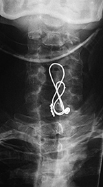
Posterior cervical wire figure of 8 construction - AP view |
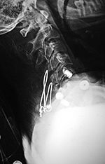
Posterior cervical wire figure of 8 construction - lateral view |
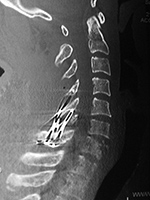
Posterior cervical wire figure of 8 construction - sagittal CT reformatted image |
 |
 |
 |
 |
| 53 year-old man with congenital cervical spine fusion and traumatic fracture through fusion mass at C3-7. The spine is stabilized by surgical fusion hardware from the occiput to T2 plus a halo brace. |
44 year-old man with C7 vertebral fracture. Posterior figure of 8 wire fixation extends from C4 to C7 |
|
| Cervical spine "corpectomy" and fusion
|
Posterior Spinal Fusion (PSF) |
 |
 |
 |
 |
| Elderly woman with diskitis at C4-5 and adjacent bony destruction by osteomyelitis at C4 and C5. Initial cervical fusion failed. A corpectomy cage was placed at C4-5 with posterior spinal fusion from the occiput to T2. A crosslink is at C6. |
57 year-old woman with rheumatoid arthritis and unstable spine. Posterior spinal fusion (PSF) is present from C3 to T2 with lateral mass screws in the cervical spine from C3-6 bilaterally and pedicle screws in the thoracic spine at T1 and T2 on the left. |
|
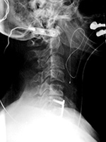
| Dorsal column stimulator, nasogastric tube, and endotracheal tube |
Nasotracheal and orgogastric tubes |
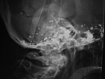
Silicone eyeball prostheses |
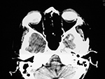
Pantopaque in the skull |
 |
 |
 |
 |
| Lateral radiograph of the neck shows a recently placed dorsal column stimulator, a nasogastric tube (black *),and an endotracheal tube (white *). |
The nasotracheal tube is larger and anterior. The orogastric tube is smaller and posterior (arrow). From Hunter, 1994 |
From Hunter, 1994 |
From Hunter, 1994 |
|
| Vagus nerve stimulator |
Esophageal temperature probe |
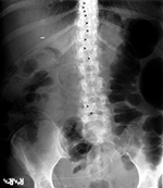
Lumboperitoneal shunt |
 |
 |
 |
 |
| A left vagus nerve stimulator was placed to treat intractable epilepsy. From Hunter, 2004 |
Also shown is an anterior cervical spine fusion plate, a monitoring electrode over an ear, and a dental bridge. From Hunter, 2004 |
45 year-old woman with two lumboperitoneal shunts to treat pseudotumor cerebri. The inferior shunt tip is at the L4-5 level (lower arrowheads). The superior shunt (upper arrowheads) goes into the thoracic region with its tip not pictured. From Hunter, 2004 |
|
| Bilateral vagus nerve stimulators
|
Dorsal column stimulation unit in upper cervical spine |
 |
 |
 |
 |
| 62 year-old man with anterior cervical fusion plate and screws C5-7 for treatment of prior cervical spine trauma. Bilateral vagus nerve stimulators were placed to treat chronic syncope.
|
From Hunter, 2004 |
|