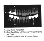
| Alloy dental restoration, root canal filling, and fixed pontic bridge with abutment crowns |
Root canals |
Dental implant crown |
Tooth numbering |
 |
 |
 |
 |
| From Harkins, 1994 |
From Harkins, 1994 |
|
The Universal/National System for permanent dentition numbers teeth beginning with the right maxillary third molar labeled as tooth number 1, following around the maxillary arch to the left maxillary third molar, descending to the left mandibular third molar, and following around the mandibular arch to the right mandibular third molar labeled tooth number 32. |
 |
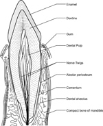
| Toot anatomy - drawing |
Tooth anatomy- radiograph |
Tooth nomenclature |
Dental overview |
 |
 |
 |
 |
| |
E - enamel; D - dentine; P - root pulp; R - tooth root; arrows - lamina dura |
WT-wisdom tooth (3rd molar); 2M-second molar; 1M-first molar; 2B- second bicuspid (premolar); 1B-first bicuspid (premolar); K9-canine; LI-lateral incisor; CI-central incisor. The * marks the mandibular canal. |
A. Alloy dental restorative; B. root canal filling with porcelain veneer crowns and posts; C. fixed bridge pontic with abutment crowns; D. dental composite (acrylic) restoration (tooth-colored fillings). From Harkins, 1994 |
|
| Miscellaneous dental devices |
Root canal fillings and porcelain denture teeth |
Cubic zirconia crown and acrylics |
Stages of dentition |
 |
 |
 |
 |
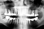
| Panoramic view of the mandible shows dental plates with screws (A) and bone ligature wires (B). There are multiple dental alloy restorations (amalgam fillings). From Harkins, 1994 |
There are root canal fillings with denture retention posts (A) and porcelain denture teeth with pins (B). From Harkins, 1994 |
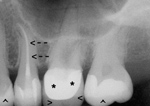
A coned mandibular view shows a cubic zirconia crown (*). The zirconia is surrounded by porcelain (horizontal arrowheads). In the next tooth there is an endodontic root canal with gutta percha (arrows), an opaque rubber. The light shade of the teeth (vertical arrowheads) shows composite acrylic restoration. |
Mixed dentition in a child with permanent teeth plus retained primary teeth. The + marks developmental follicles for the third molar and the * marks permanent second molars. |
|
| Removable partial denture framework (A); dental crowns (caps) (B) |
Temporomandibular prosthetic condyle implant (A); orthodontic arch bars (B); porcelain veneer dental crowns (caps) (C); fixation screws (bone screws) (D) |
Osseointegrated dental implants with fixed dental bridgework (A); maxillary denture with acrylic teeth (B) |
Subperiosteal dental implant with fixed bridge (A); bone plate with screws (B); fixed dental bridgework with root canal fillings (C); fixation wire (D) |
 |
 |
 |
 |
| From Harkins, 1994 |
|
| Dentures (false teeth) |
|
|
 |
 |
|
|
| 78 year-old woman with sinus headaches. Incidental presence of dentures (false teeth). Note air-fluid levels in both maxillary sinuses. |
|
|
|
| Zimmer Trabecular Metal Implant |
Zimmer Trabecular Metal Implant (on left) and conventional metal implant (on right) |
Root canal |
_small.jpg) |
 |
 |
 |
| |
|
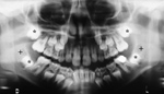
Periodontal abscess around the roots of the right first molar (arrows) in a 67-year old man. |
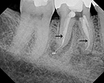
Periodontal abscess after root canal treatment. The arrows point to the opaque sealant used to seal the cleaned out root canal. |
|
| Orthodontic appliance (braces) |
Dental caries, mandibular trauma, dental amalgam, and lip ornaments |
Permanent mandibular retainer |
Fibular mandibular reconstruction |
 |
 |
 |
 |
| Adolescent male who was in an altercation sustaining a fracture of the right mandibular angle and the left mandibular body. The braces are unrelated to the trauma. |
Frequent emergency department visitor with bilateral mandible fractures, scattered dental caries, impacted right mandibular wisdom tooth, dental amalgam, a right maxillary root canal, and upper and lower lip ornaments. |
Panoramic view of 38 year-old woman with a permanent (metallic) mandibular retainer. |
25 year-old woman with mandibular osteosarcoma. The mandible was restored with autologous fibula free-flap reconstruction. |


.jpg)


