| Right shoulder hemiarthroplasty anterior dislocation |
Right shoulder prosthesis periprosthetic fracture |
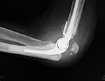
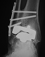
Left elbow semiconstrained prosthesis with olecranon periprosthetic fracture |
 |
 |
 |
 |
| |
|
51 year-old woman. From Benjamin, 1994 |
|
 |
| Right shoulder hemiarthroplasty failure with loosening and osteolysis |
|
 |
 |
 |
|
| 53 year-old man in motorcycle accident. Initial radiograph shows a comminuted fracture of the proximal right humerus with dislocation of the humeral head (left image). The fracture was treated with a right shoulder hemiarthroplasty (center image). At follow-up 6 months later there was failure of fracture healing with associated hardware loosening and osteolysis (arows on right image). Images courtesy Laura H Lee, MD. |
|
|
| Posterior dislocation of a reverse total shoulder arthroplasty |
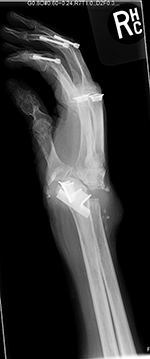
Disrupted wrist prosthesis |
Second generation total wrist arthroplasty and Silastic metacarpophalangeal (MCP) implants. |
 |
 |
 |
 |
| |
|
Elderly woman with advanced rheumatoid arthritis. Swanson silicone joint prostheses are present at the right 1st, 2nd, and 3rd metacarpophalangeal joints, and Herbert-like screws are at the proximal interphalangeal joints of the right 2nd, 3rd, and 4th fingers. The Swanson silicone prosthesis at the wrist radiocarpal joint has become completely disrupted with displacement of the radial component. |
The wrist is misaligned in ulnar deviation. There is extensive remodeling in the long (3rd) finger metacarpal. Metal grommets with Silastic implants are at the thumb and long finger MCP joints. From Hunter, 1994. |
 |
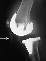
| Right bipolar radial head prosthesis with eventual prosthesis loosening |
 |
 |
 |
 |
| 57 year-old woman with radial head prosthesis and capitellar suture anchor. In 2011 she had a right elbow dislocation with a comminuted radial head fracture. A bipolar radial head prosthesis was placed to treat post-traumatic arthritis. Initial radiographs obtained in 2014 (left two images) showed the prosthesis to be well seated. Later radiographs in 2015 show prominent periprosthetic lucency around the prosthetic stem indicating non-infectious aseptic loosening. There are also chronic fractures of the proximal portions of the right radius and ulna. |
|
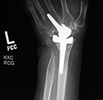
| Third generation "biaxial" total wrist prosthesis |
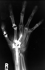
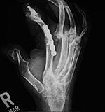
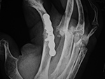
Multiple failed metacarpophalangeal Swanson silicone arthroplasties |
 |
 |
 |
 |
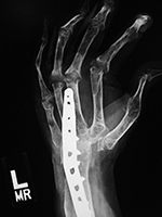
| 52 year-old woman with rheumatoid arthritis and left total wrist prosthesis which failed three years later and was replaced by a wrist arthrodesis. The distal stem of the prosthesis protruded out of the left 3rd metacarpal. The radiograph of the left wrist arthrodesis shows 2nd-5th MCP Swanson silicone arthroplasties, most of which are at least partially displaced. On the patient's right side there are also extensive changes of rheumatoid arthritis with multiple failed Swanson silicone MCP prostheses (see the two images to the right). |
52 year-old woman with severe rheumatoid arthritis in the right hand and wrist. There are multiple failed MCP Swanson silicone prostheses with partial displacement and bone fracturing. There is surgical arthrodesis at the right index finger MCP joint and bony fusion from long standing arthritis in the wrist and thumb MCP joint. In her left hand and wrist there was failure of a wrist prosthesis and failure of MCP joint arthroplasties (see the two images to the left). |
|
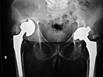
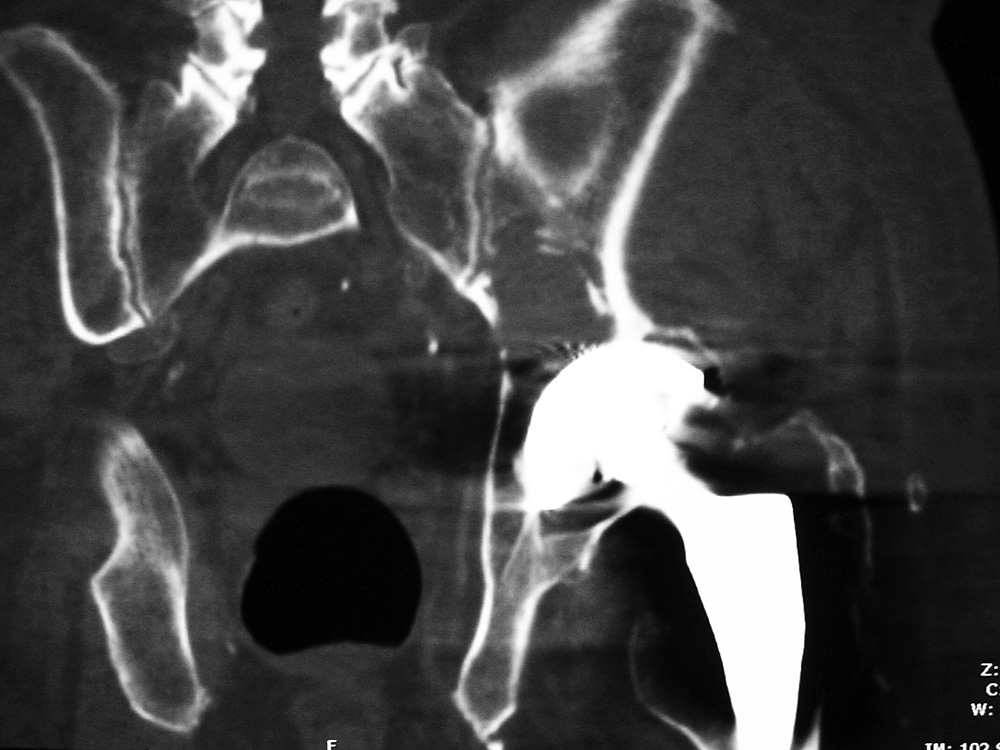
| Osteolysis and particle disease in right hip replacement |
Osteolysis and particle disease in right hip implant
|
 |
 |
 |
 |
|
|
There is displacement of the right acetabular implant component and osteolysis from granulomatous particle disease. A left metal upon metal hip implant is present. |
| Left hip arthroplasty: polyethylene liner wear with associated osteolysis and particle disease |
 |
 |
 |
 |
|
|
Axial CT image |
Coronal CT image |
|
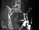
| Particle disease (arrows) left hip coronal CT image |
Focal osteolysis about femoral stem of total hip arthroplasty |
Femoral osteolysis and loosening of noncemented total hip arthroplasty |
Right ankle prosthesis failure with loosening of components and polyethylene wear medially |
 |
 |
 |
 |
| 68 year-old man with particle disease from a worn out left hip prosthesis. Bony destruction (arrows) is in the left supra-acetabular region and in the left greater trochanter with a pathologic fracture. |
Cemented total hip arthroplasty with focal osteolysis about the stem in zones, 2, 5, and 7. From Hunter, 1994. |
There is radiolucency (arrow) around the entire femoral stem with a sclerotic margin. There is also thinning of the lateral femoral cortex. From Hunter, 1994. |
68 year-old woman with total ankle prosthesis failure with loosening of tibial and talar component shown by bony lucency about the components and medial polyethylene liner wearing. |
|
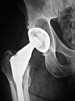
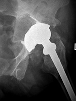
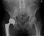
| Right hip arthroplasty polyethylene wear |
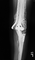
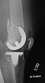
Left total knee prosthesis patellar button dislocation |
Right total knee prosthesis patellar button dislocation |
 |
 |
 |
 |
|
The patellar button is dislocated superiorly (arrow). |
The patellar component (button) is dislocated inferiorly (arrow) |
|
| Displaced polyethylene liner in right hip arthroplasty |
Semiconstrained left elbow arthroplasty with heterotopic bone formation |
|
 |
 |
 |
|
| Image courtesy Lana Hirai Gimber, MD |
35 year-old woman with prior gunshot injury to left upper extremity. Multiple surgeries were performed to remove wooden fragments and shrapnel. Extensive heterotopic bone has developed about the left humerus and left elbow arthroplasty. Portions of previous fixation screws and plates are evident. |
|
|
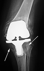
| Disrupted left total knee prosthesis |
Failed constrained total knee arthroplasty from destructive particle disease |
 |
 |
 |
 |
| |
|
96 year-old woman with destructive particle disease causing large areas of bone destruction (arrows). |
|
| Right femur supracondylar periprosthetic fracture |
 |
 |
 |
 |
| 74 year-old women who was in an automobile accident and suffered a right femur supracondylar periprosthetic fracture about her cruciate retaining TKA. The fracture was treated with a lateral periarticular plate and screws. |


