Orthopedic Devices
|
|
|
|
Internal Fixation - nails and rods |
|
|
|
|
|
|

|
Fracture Fixation
by Tim B Hunter, MD, MSc
Internal Fixation continued
Intramedullary Nails/Rods
Numerous intramedullary nails or rods of different
design are available (figure: intramedullary rods and nails). Femoral nails
are bowed anteriorly to accommodate the contour
of the femur. A majority of nails are cannulated to allow their placement over a guide wire. Intramedullary
nails provide excellent stability
against bending forces, but they do not control
rotation and compressive forces. For the control
of rotational forces, proximal and distal interlocking
screws are placed (usually in a lateral to medial
fashion) through the nail or rod holes in the
proximal and distal femur. Interlocking screws
increase fixation stability and therefore led to an
increased use of nailing in fracture fixation.
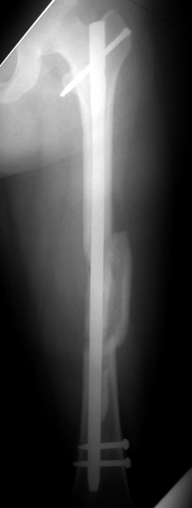
Short hip or femoral nails are often used with proximal femoral fractures, especially intertrochanteric fractures. These nails are thicker and more rigid to withstand the stress in the intertrochanteric and subtrochanteric regions. They have an accompanying femoral neck screw of standard design or of helical (spiral design) (figure: short hip nail). The
weakest points of these nails are
the distal interlocking screws. In children
with osteogenesis imperfecta, two-part telescoping
rods are used to allow lengthening of the
rod as the child grows (Benjamin, 1994).
Interlocking
(locking) screws were introduced by Grosse and
Kemp with one to two proximal
and usually two distal interlocking
screws in femoral nails and three in tibial
nails. The proximal femoral screws can be placed
either (more commonly) obliquely through the intertrochanteric
region or perpendicular through
the proximal femoral shaft. The distal interlocking
screws are placed perpendicular to the distal
femoral shaft (figure: blade spiral distal locking screw). Interlocking screws also prevent
collapse or shortening of the fracture (Ruedi, 2007).
If a nail is locked both proximally and distally,
it is statically locked because all planes of
motion are controlled or static. A nail is dynamically
locked if it is locked at one end only,
which allows compression at the fracture site. Dynamization produces increased compression
at the fracture site after the nail is unlocked at one
end by removal of the interlocking screws. It is
rarely needed in the femur, but it may be recommended
in the tibia for certain fracture patterns (figure: tibia dynamization).
Dynamization is usually performed 2–3 months
after initial surgery when one or both proximal
interlocking screws are removed. Because unreamed
nails are thinner, the use of interlocking
screws is mandatory with unreamed nails to prevent torsion (Ruedi, 2007; Benjamin, 1994; Hunter, 2001). Reconstruction nails have been designed for
the treatment of femoral shaft fractures with ipsilateral
femoral neck, intertrochanteric, or subtrochanteric
fractures. These nails have proximal
locking holes oriented to accommodate screw
placement into the femoral neck and head (figure: intramedullary rods and nails).
Flexible intramedullary rods are of smaller diameter
and greater flexibility than standard rods and nails to accommodate
different variations in long bone anatomy (Enders nail; Lottes nail; and Rush pin) (figure: Enders (flexible) fixation nails; figure: Rush pin). These nails are solid
and are associated with a lower prevalence of infection
than the larger cannulated rods. Flexible rods
are inserted through the metaphysis. They are
frequently used for fixation of long bone diaphyseal
fractures in skeletally immature patients to
avoid placement through the growth plate and
subsequent premature closure of the growth
plate. Multiple flexible rods, which diverge in the metaphyseal regions, are placed through multiple
insertion sites. These rods provide some axial and
rotational stability. For small-diameter bones,
sometimes a single flexible rod is used. The major
disadvantage associated with flexible rods is the common need for additional external fixation, such as a plaster cast (Ruedi, 2007; Benjamin, 1994; Berquist, 1995; Hunter, 2001; Oh, 2002).
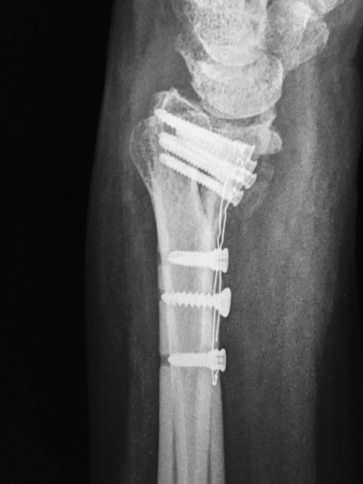
Rush pins are thin fixation rods with a sled-runner tip and a hooked end. They are frequently employed for treatment of ulnar shaft fractures or fractures in other long thin bones where intramedullary insertion of the Rush pin can reduce and stabilize a fracture (figure: ulnar Rush pin). A Rush rod is similar with a chisel like tip. It is typically used in fibular shaft fractures (figure: fibular Rush rod).
Back to Top
Back to Top
|