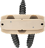
| Harms vertebral cage (AP view) |
Harms vertebral cage (lateral view) |
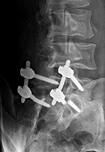
Lumbar spine bony disk strut, pedicle screws, and pedicle rods (AP view) |
Lumbar spine bony disk strut, pedicle screws, and pedicle rods (lateral view) |
 |
 |
 |
 |
| There is a vertebral cage and side plate and screws in the lower thoracic spine for treatment of a spinal tumor. From Hunter, 1994 |
From Hunter, 2004 |
 |
| Vertebral corpectomy with vertebral cage and left lateral side plate |
 |
 |
 |
 |
| 70 year-old man with history of L1 and L3 injury and T11-L3 spinal fusion in the 1990's. Recent worsening of chronic lower back pain. Standard radiographs (left two images) show a vertebral body corpectomy cage at T12-L3 with placement of a left lateral side plate at the same levels. There are two proximal screws at T12, one of which enters the spinal canal as shown on subsequent CT (right two images). There are two distal screws at L3, the most distal of which enters the L3-4 disk space (lateral radiograph). |
 |
| Vertebral compression fracture treated by expandable corpectomy device |
TLSO brace (Boston brace) |
 |
 |
 |
 |
| |
There are also bilateral pedicle screws and connecting rods above and below the level of the fracture. |
The brace stabilizes an L1 compression fracture. |
 |
| Anterior lumbar interbody fusion (ALIF) |
Pedicle fixation screws and rods |
 |
 |
 |
 |
| 47 year-old woman with anterior lumbar interbody fusion (ALIF) at L5-S1. |
20 year-old woman with L1 vertebral body compression fracture treated with T12-L2 posterior spinal fusion using pedicle screws at T12 and L2 with connecting rods on each side. |
 |
| Anterior lumbar interbody fusion (ALIF) |
ILIF: interlaminar lumbar instrument fusion |
 |
 |
 |
 |
| 60 year-old man with L5-S1 anterior diskectomy and interbody fusion with zero-profile fixation screws at L5-S1. There are PEEK interbody disk grafts at multiple levels from XLIF prior surgery at L2-3 to L4-5. |
67 year-old woman with L3-4-5 ILIF. There are partial laminectomies at L3 and L4. Posterior L3-5 stabilization is obtained with a spinous process clamp. There is also posterior L3-5 arthrodesis using structural allograft. The patient is wearing a TLSO brace. |
 |
| Interspinous clamps (ILIF) at L4-5 |
Interspinous clamps (ILIF) at L4-5 |
 |
 |
 |
 |
| 67 year-old woman with lower lumbar spinal stenosis. The patient is wearing a TLSO brace. |
76 year-old woman treated for degenerative spondylolisthesis at L4-5. The patient is wearing a TLSO brace. |
 |
| Posterior Instrumented Lumbar fusion (PLIF) |
X-stop device |
 |
 |
 |
 |
| Screws are placed through the pedicles into the vertebral bodies. The screws are connected together on each side with rods or a plate placed over the pedicle screws on each side. Some of these systems are also combined with posterolateral bony fusion masses. |
Image from rheumatology network |
 |
| X-Stop Devices |
|
 |
 |
 |
|
| 69 year old man with chronic low back pain treated with X-Stop devices at L4-5 and L5-S1. Sagittal T1-w MRI (3rd image) shows some metallic susceptibility artifact but the spinal canal is visible. |
|
 |
| Steffee Plates - Posterior Instrumented Lumbar Fusion (PLIF) |
Coflex device |
 |
 |
 |
 |
| There is also a Brantigan disk cage at L5-S1 (arrow on lateral view) |
The Coflex device is made from titanium. The "U" shaped device is positioned horizontally with its apex anterior. The two long arms of the "U" parallel the long axis of the spinous process. The ridges help keep the device stably attached to the spinous process. Image from Paradigm Spine |
 |
| ProDisc L |
Lumbar disk replacement |
 |
 |
 |
 |
| ProDisc L (Synthes) artificial lumbar disk. © DePuy Synthes 2016. All rights reserved. ProDisc® L is a trademark of DePuy Synthes. |
There is also posterior spinal fusion. |
 |
| PEEK cages packed with iliac crest autograft material in anterior lumbar instrumented fusion (ALIF) with posterior spinal fusion (PSF) at L5-S1 |
 |
 |
 |
 |
| 48-year old man with L5-S1 laminectomy and posterior spinal fusion (PSF) for radiculopathy and isthmic spondylolisthesis. There was hardware failure. A subsequent ALIF with a Sovereign cage device (Medtronic) was performed as well as revision of the PSF at L5-S1. Image in the fourth column Reprinted with the permission of Medtronic, Inc. © 2016 |
 |
| Occiput-T8 posterior fusion with anterior cervical fusion from C6-T1. |
 |
 |
 |
 |
| 59 year-old man with occiput to T8 posterior fusion. There is anterior cervical fusion from C6-T1. PEEK disk cages are present at C6-7 and C7-T1. An old compression fracture is present at T5. In the cervical spine lateral mass screws are at C3-6 bilaterally. In the thoracic spine pedicle screws are at T1-3 and T6-8 on the right and at T1-4 and T6-8 on the left. Laminectomies have been performed from C3 to C6. |