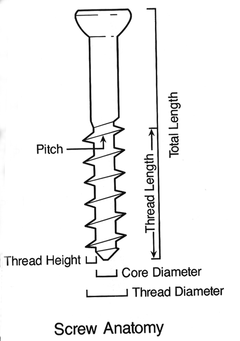
| The anatomy of a fixation screw |
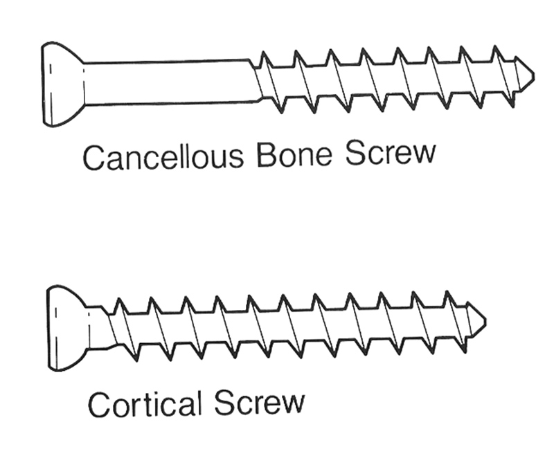
Cancellous and cortical bone screws |
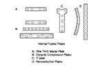
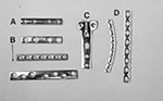
Drawing of fracture fixation plates |
Fixation plates |
 |
 |
 |
 |
| From Hunter, 1994 |
From Hunter, 1994 |
From Hunter, 1994 |
A - one third tubular plate; B - dynamic compression plate (DCP); C- T-plate; D - reconstruction plates. From Hunter, 1994 |
 |
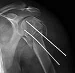
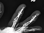
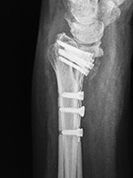
| K-wires AP view |
K-wires lateral view |
Herbert screw |
Herbert screws |
 |
 |
 |
 |
| 50 year-old woman with a distal radius immobilized by 4 Kirschner wires (K-wires). There is also an ulnar styloid fracture. |
Note the different screw pitch on each end. Also known as headless (recessed) fixation screws. From Taljanovic, 2005 |
 |
| Herbert screw |
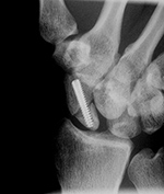
Acutrak screw |
Steinman pins |
Rush pin (ulna) and dynamic compression plate (radius) |
 |
 |
 |
 |
| There is a healing scaphoid fracture, and a fiberglass cast is in place. From Hunter, 1994 |
The Acutrak screw stabilizes a scaphoid fracture. |
From Taljanovic, 2005 |
|
 |
| Olecranon plate and screws plus radial head prosthesis |
 |
 |
 |
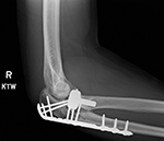 |
| 35 year-old man who fell and sustained comminuted olecranon and radial head fractures. He was treated with olecranon plate and screws as well as a radial head prosthesis. Courtesy Lana Hirai Gimber, MD, MPH |
|
| Shoulder capsule anchor fixation screws |
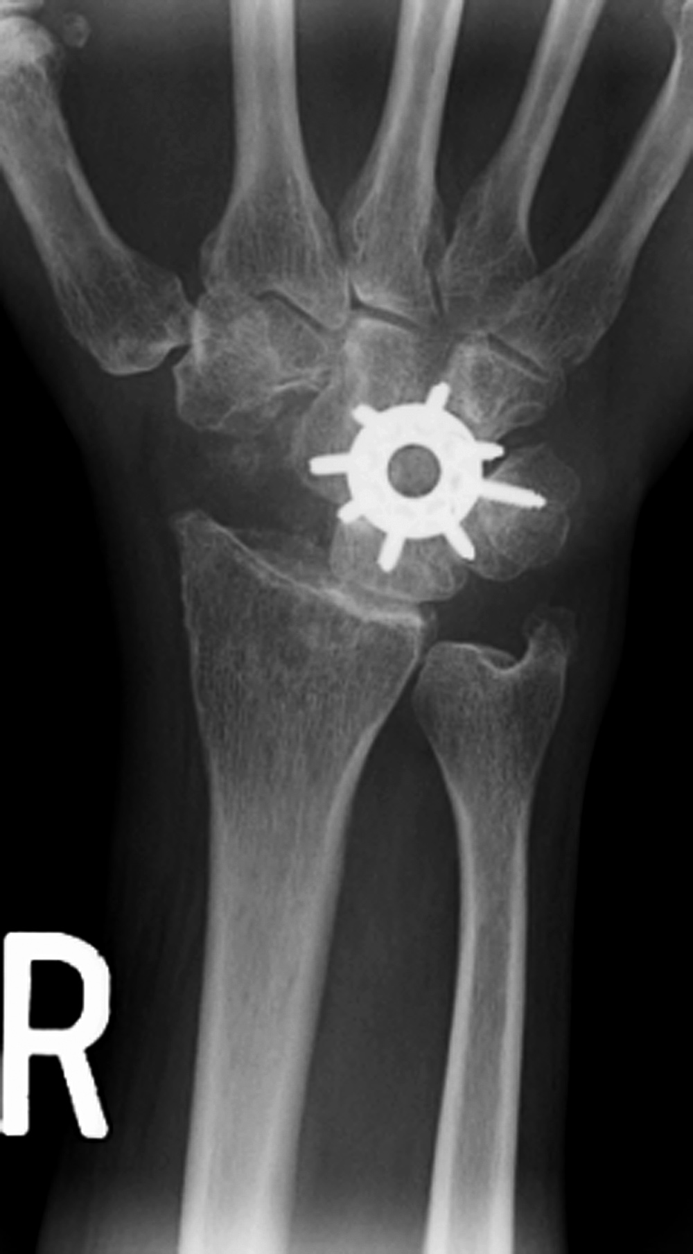
Spider plate |
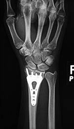
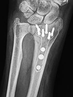
Variable angle volar distal radius locking plate |
 |
 |
 |
 |
| |
The scaphoid has been removed. The spider plate transfixes the lunate, triquetrum, capitate, and hamate which are partially fused. From Taljanovic, 2005 |
20 year-old woman with surgical plate placement for a malunited distal radius fracture. From Taljanovic, 2005 |
|
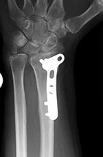
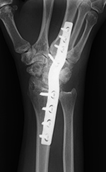
| Wrist T-plate (AP view) |
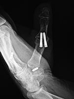
Wrist T-plate (lateral view) |
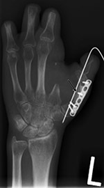
Acutrak screws and endobuttons |
 |
 |
 |
 |
| This is also commonly called a "volar" fixation plate. From Taljanovic, 2005 |
64 year-old woman with thumb "tightrope" surgery using Endobuttons and Acutrak screws. The trapezium has been resected. |
|
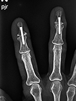
| Acutrak screws |
Little finger reconstruction plate |
Interference (Kurosaka) screw |
 |
 |
 |

|
| The Accutrak screws are used for arthrodesis of the distal interphalangeal joints of the right index and long fingers in a woman with severe osteoarthritis. |
47 year-old man with fracture of the left little finger proximal phalanx. |
Interference (Kurosaka) screws are used to anchor anterior cruciate ligament graft in femoral and tibial metaphysis. Note the multiple osteochondromas (familial exostosis). From Hunter, 1994 |
|
| Suture anchors (anchor screws) in humeral head |
Low contact dynamic compression plate used for wrist arthrodesis |
Short reconstruction plate and Kirschner wire |
 |
 |
 |
 |
| Multiple suture anchors are present in the left shoulder humeral head for tacking down the joint capsule in a patient with recurrent shoulder dislocation. |
From Taljanovic, 2005 |
The arrow points to bone graft at osteotomy site. |
|
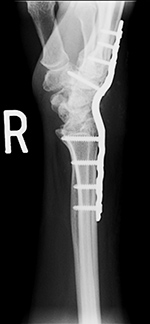
| Locking dynamic compression plate (LCD) and screws transfix a healed distal radius shaft fracture |
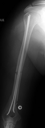
Enders (flexible) fixation nails |
Flexible fixation rods |
 |
 |
 |
 |
| There is also a volar periarticular fixed angle plate and screws with a proximal locking screw that transfixes a new healing distal radial diametaphyseal fracture. From Taljanovic, 2005 |
From Taljanovic, 2005 |
Flexible intramedullary fixation rods stabilize a healing humerus shaft fracture. The wire in the eyelets distally (arrow) prevent the rods from backing out of the insertion portal. From Hunter, 1994 |
|
| Tension band wiring |
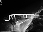
Clavicular hooked plate |
Clavicle reconstruction plate |
Carbon fiber humerus fixation plate |
 |
 |
 |
 |
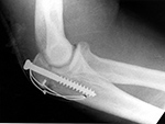
| Tension band fixation of an olecranon fracture using a cancellous screw. The looped wire transforms the distractive pull of the triceps into compression at the fracture site. From Hunter, 1994 |
|
|
The carbon fiber fixation plate itself is radiolucent but has a wire encased in it to allow radiographic evaluation of its placement. In the proximal end of the plate are 3.5 mm proximal humeral head fixed angle threaded locking holes with 3.5 mm locking screws.
Image courtesy Brandon Runyan, MD. |
|
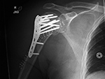
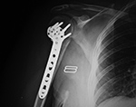
| Bilateral proximal humerus periarticular locking plates |
Carbon fiber radius volar fixation plate |
 |
 |
 |
 |
| 57 year-old man with comminuted bilateral proximal humerus fractures. Skin staples are present. There are old, healed rib fractures on the left. |
The carbon fiber fixation plate itself is radiolucent but has a wire encased in it to allow radiographic evaluation of its placement.
Image courtesy Brandon Runyan, MD. |
|
| Periarticular locking plate |
Humerus periarticular locking plate with Vitoss bone substitute |
Multiple humeral head suture anchors
|
 |
 |
 |
 |
| |
|
60 year-old woman with repair of a massive rotator cuff tear. Three suture anchors are present. There are old, healed left rib fractures. |