|
|

|
Dental Devices
by Tim B Hunter, MD, MSc and Rick Light, DDS
Tooth Anatomy
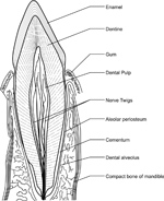
A thorough knowledge of tooth anatomy is important for proper diagnosis of dental disease and for recognition of dental apparatus. Each tooth contains distinct layers visible on conventional radiographs (figure: tooth anatomy). The anatomic crown is the portion of the tooth that is covered by enamel. Enamel, formed from ameloblasts, is the most dense tissue in the human body, composed of 97% inorganic salts. Enamel appears as the most radiopaque layer of the tooth on conventional radiographs.
The layer deep to the enamel, dentin, is a calcified, porous tissue, the second most dense tissue in the body. Dentin, laid down by odontoblasts, is a comprised of 65% inorganic salts, and forms the bulk of the tooth. Dentine is less dense than enamel, and appears as a gray opacity on conventional radiographs (figure: tooth anatomy). Dentin surrounds the pulp chamber, which lies deep within the anatomical crown, and the pulp, or root canals within the root of the tooth. Pulp consists of connective tissue, nerves, lymph channels, and blood vessels and is visualized as a radiolucency generally in the center of the anatomical crown and root extending to the apex. Reserve odontoblasts embedded within the lacunae of the dentin lay down secondary, or reparative dentin and reduce the pulp cavity and root canal size with age.
The tooth root is the elongated internal structure of the tooth and begins where the enamel covering ends. This junction is called the cemento-enamel junction (CEJ). The alveolar bone socket begins approximately 1.8 mm from the CEJ. The non-covered root distance is called the biologic width and is required for gingival, or epithelial attachment. Alveolar bone surrounds the roots and supports the tooth. If a tooth is extracted, the alveolar bone will in time be resorbed. Pathological processes, such as periodontal disease, can remove alveolar bone as well. Alveolar bone loss should be suspected when the distance from the alveolar ridge to the cemento-enamel junction of the supported tooth is greater than 1.8 mm, or when the opacity of the alveolar crest is lost. Below the alveolar bone is basal bone. The transition between the two is not radiographically visible.
In an individual with either advanced, chronic, or refractive periodontal disease alveolar bone may be completely lost leaving only the residual basal bone. The wall of the alveolar socket, or lamina dura, is made of dense cortical bone. It appears on a radiograph as a white line next to the dark line of the periodontal ligament space. The periodontal ligament space, containing the periodontal ligament, is visualized between the lamina dura and the tooth root as a radiolucent line.
The periodontal ligament is comprised of radiolucent collagen bundles and functions to stabilize the teeth within their sockets. Its appearance varies from tooth to tooth, depending upon the overall periodontal health. It generally has a thin radiolucent appearance from the CEJ to the root apex. Endodontic or dental pulp pathology shows as a widened radiolucency within the periapical region that may extend toward the top of the tooth but narrows as it goes superiorly. Periodontal pathology, on the other hand, shows as an ever widening radiolucency from the tooth root toward the CEJ and the top of the tooth.
Cementum is a bone like substance forming the thin surface layer over the tooth roots. It functions as an anchor point for the attachment of periodontal ligaments. Cementum on the root surface is nearly the same density as the dentin, thus it is usually not visible radiographically.
Teeth are a combination of living tissues and non-living structures. The living tissues are the cementum and pulp. The non-living structures are the dentin and enamel.
Stages of Dentition
Three separate stages of dentition occur in humans (figure: stages of dentition). The earliest stage lasts up until approximately six years of age with the mouth containing 20 temporary deciduous teeth. During the second stage from ages 6 to 21, both the deciduous temporary teeth and the permanent teeth replacing them are present in the oral cavity. The timing of permanent tooth eruption varies according to the specific tooth. The third and final stage of dentition is observed in the adult. Commonly, this stage demonstrates 32 teeth, containing three molars, two premolars, one cuspid, and two incisors in each of the four quadrants in the mouth. Although 20 deciduous teeth and 32 permanent teeth is the most common presentation, patients may vary in their number of teeth. Adontia is the developmental absence of teeth, and may be total or partial. Extra teeth are a more common variant, particularly in the incisor region, and are referred to as supernumerary teeth.
It is not uncommon to see dental braces during adolescence (figure: braces). Typically, all the permanent teeth are in place by then. Braces have traditionally been used for cosmetic dental purposes in adolescents and young adults, but they have many other applications and many differing designs. They align and straighten teeth and can be applied to correct underbites, malocclusions, overbites, and crooked teeth, as well as addressing other congenital or traumatic flaws in the teeth and jaws.
Tooth Nomenclature
Tooth nomenclature and numbering are important for properly describing dental and mandibular lesions. Tooth nomenclature is based on tooth location and anatomic features (figure: tooth nomenclature). Central teeth refer to the two upper and two lower teeth in the most mesial, or medial, position of the mouth. Lateral teeth are the teeth directly lateral to the centrals. The centrals and laterals are also collectively referred to as the incisors. Cuspid teeth, or canines, are just lateral and posterior to the laterals and have only one cuspal or point. Bicuspids, or premolars, are located posterior to the cuspids and have two cuspals (points). Molars are posterior to the bicuspids and usually contain four cusps. The deciduous arch has four molars and no bicuspids. The permanent arch contains four bicuspids and six molars (figure: tooth nomenclature).
Numbering of the teeth is important for consistent description of dental anatomy. The American Dental Association recognizes two major systems for numbering the teeth. The Universal/National System is used primarily in the United States, and the International Standards Organization System (ISO) is used in most other countries. The Universal/National System for permanent dentition numbers teeth beginning with the right maxillary third molar labeled as tooth number 1, following around the maxillary arch to the left maxillary third molar, descending to the left mandibular third molar, and following around the mandibular arch to the right mandibular third molar labeled tooth number 32 (figure: tooth numbering).
The Universal/National System order for the primary (baby) dentition is the same as described for the permanent dentition; however, the primary teeth are designated by upper case letters A through T, with A being the patient's right maxillary second primary molar and T being the right mandibular second primary molar.
The World Dental Federation (FDI) ISO notation divides dentition into quadrants. In this system, the quadrants and teeth are designated by sequentially moving right to left and down across the oral cavity. Permanent dentition quadrants are numbered as follows: 1-right upper maxilla, 2-left upper maxilla, 3-left lower mandible, 4- right lower mandible. Deciduous quadrants are numbered 5-right upper maxilla, 6-left upper maxilla, 7-left lower mandible, 8- right lower mandible. The beginning point in the nomenclature of the teeth within each quadrant is the midline. Therefore, the upper right adult central incisor would be notated as 11 (replacing the deciduous central incisor notated as 51), while the lower left adult second bicuspid would be notated as 35 (replacing the deciduous second molar notated as 75).
Tooth Anatomy
| Tooth anatomy |
Stages of dentition |
Tooth nomenclature |
 |
 |
 |
 |
| E - enamel; D - dentine; P - root pulp; R - tooth root; arrows - lamina dura |
Mixed dentition in a child with permanent teeth plus retained primary teeth. The + marks developmental follicles for the third molar and the * marks permanent second molars. |
WT-wisdom tooth (3rd molar); 2M-second molar; 1M-first molar; 2B- second bicuspid (premolar); 1B-first bicuspid (premolar); K9-canine; LI-lateral incisor; CI-central incisor. The * marks the mandibular canal.
|
 |
| Tooth Numbering |
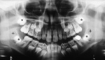
Dental caries, mandibular trauma, dental amalgam, and lip ornaments |
Root canal |
 |
 |
 |
 |
| The Universal/National System for permanent dentition numbers teeth beginning with the right maxillary third molar labeled as tooth number 1, following around the maxillary arch to the left maxillary third molar, descending to the left mandibular third molar, and following around the mandibular arch to the right mandibular third molar labeled tooth number 32.
|
Frequent emergency department visitor with bilateral mandible fractures, scattered dental caries, impacted right mandibular wisdom tooth, dental amalgam, a right maxillary root canal, and upper and lower lip ornaments. |
Periodontal abscess around the roots of the right first molar (arrows) in a 67-year old man. |
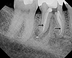
Periodontal abscess after root canal treatment. The arrows point to the opaque sealant used to seal the cleaned out root canal. |
Back to Top
|


