There are many kinds of cervical spine immobilization devices, including cranial (head) tongs (figure: cranial tongs), cervical collars, neck braces, and halo vests. Cervical collars are ubiquitous and are commonly placed on trauma patients in the emergency department. The most frequent cervical collar design is the Philadelphia collar, which is molded from plastic and has chin and occipital supports. Although it is effective in stabilizing the neck to prevent harmful motion, it is uncomfortable, and patients want to remove it as soon as possible.
A more comfortable collar for patients is a soft foam collar covered by cotton. Although this collar is ineffective in controlling neck motion, it is useful as a reminder to the patient that neck motion must be avoided.
No neck collar provides adequate long-term neck stabilization for unstable cervical spine fractures. Unlike cervical collars, cervical braces are designed for the long-term treatment of cervical spine fractures. They consist of chin and occipital supports that are connected to a thoracic vest by metal rods. Cervical braces do provide good prevention against harmful flexion, but they are not as effective in preventing harmful extension.
| Cervical spine disk cages |
Cervical spine "corpectomy" and fusion |
 |
 |
 |
 |
| 65 year-old woman with vertebral disk cages at C3-4, C4-5, and C5-6. A previous anterior C4-6 fusion plate has been removed. The disk cages are probably composed of PEEK. |
Elderly woman with diskitis at C4-5 and adjacent bony destruction by osteomyelitis at C4 and C5. Initial cervical fusion failed. A corpectomy cage was placed at C4-5 with posterior spinal fusion from the occiput to T2. A crosslink is at C6. |
 |
| Cervical spine PEEK disk cages |
Occipital strut and posterior cervical spine fixation for C1-2 subluxation |
Zero-profile ACDF |
 |
 |
 |
 |
| 56 year-old man with severe spinal stenosis. There are PEEK disk cages at C3-4, C4-5, C5-6, and C6-7 held in place by anterior interbody screws as well as bony anterior cervical fusion. A feeding tube is also partially visualized. |
Middle-aged woman with rheumatoid arthritis and unstable atlantoaxial (C1-2) subluxation. Note the wide separation between the odontoid and the anterior ring of C1. The cervical spine was stabilized with an occipital strut held in place by occipital screws and rods extending from the occiput to C2. Lateral mass screws are present connecting to the rods at C2, and there is also posterior spinal wiring. |
54 year-old woman with C6-7 zero profile ACDF discovered incidentally on a thoracic spine series after a fall. A PEEK disk cage is at C6-7 held in place by anterior interbody screws. |
|
| Posterior cervical spine fusion from occiput with halo brace |
Posterior cervical wire figure of 8 construction - AP view |
Posterior cervical wire figure of 8 construction - lateral view |
Posterior cervical wire figure of 8 construction - sagittal CT reformatted image |
 |
 |
 |
 |
| 53 year-old man with congenital cervical spine fusion and traumatic fracture through fusion mass at C3-7. The spine is stabilized by surgical fusion hardware from the occiput to T2 plus a halo brace. |
44 year-old man with C7 vertebral fracture. Posterior figure of 8 wire fixation extends from C4 to C7 |
|
| Cranial (head) tongs |
Cranial (head) tongs |
 |
 |
 |
 |
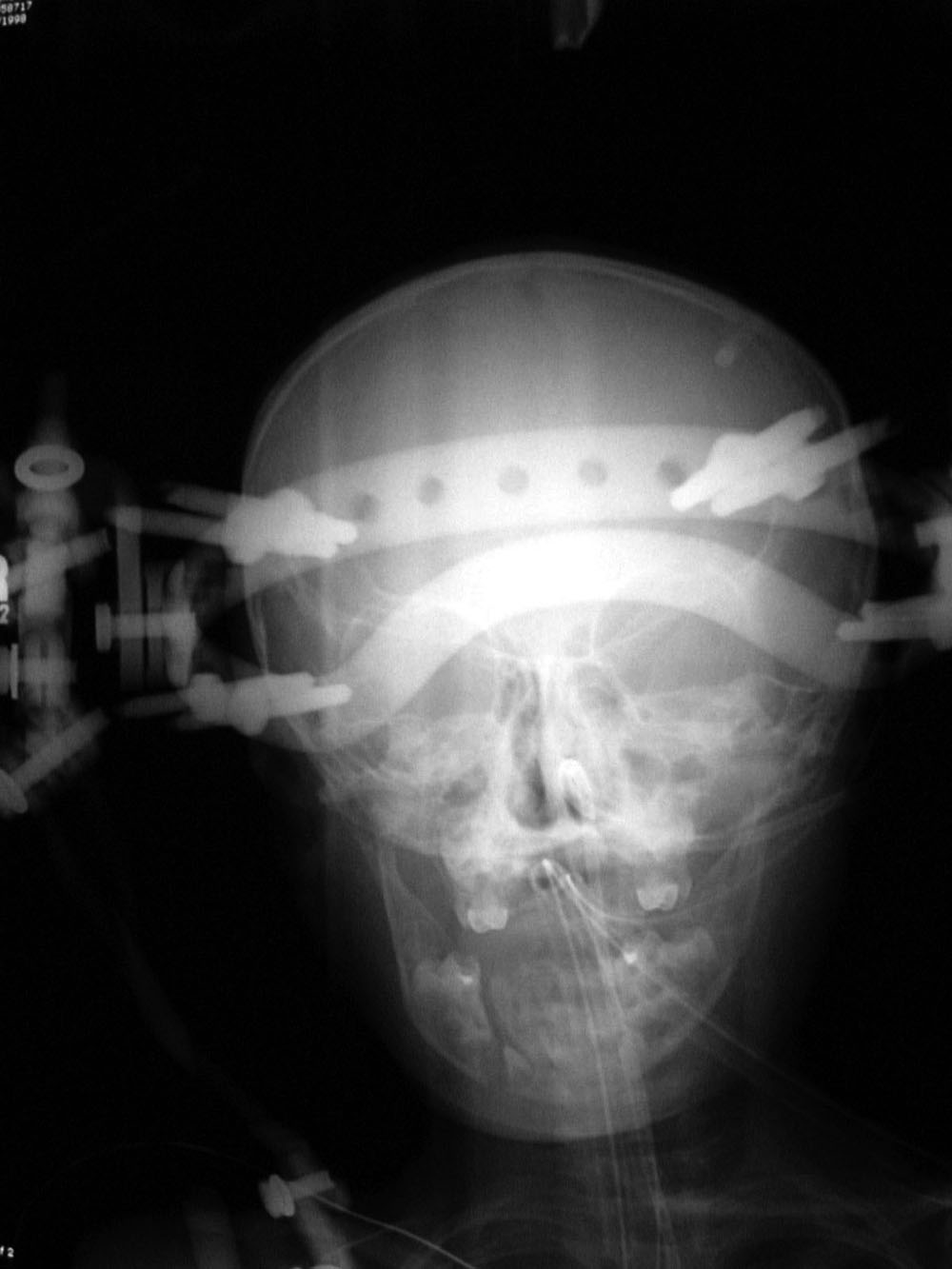
| Child with severe intracranial and cervical spine injuries with bilateral cranial stabilization tongs, an endotracheal tube, an oroogastric tube, and a feeding tube entering via the nose. From Hunter, 2004 |
Cranial (head) tongs are used to stabilize the head and neck in a patient with a cervical spine fracture. One or more screws penetrate the outer table of the skull on each side. They are connected to each other by horizontal or vertical bars on each side that are attached to an external traction device. From Hunter, 2004 |
|
| Posterior cervical spine wiring |
Halo vest and brace |
 |
 |
 |
 |
| Posterior cervical wires from C4 to C7 after laminectomy. The lateral view obtained after cervical myelography. From Yoshino, 1994 |
From Yoshino, 1994 |